
Anatomy of Flowering Plants Notes
Study Notes
Topics
9Chapter Overview, Core Logic and Mind Map
Overview
Anatomy of flowering plants explains the internal organisation of roots, stems and leaves by studying tissues and tissue systems. NCERT first divides plant tissues into meristematic tissues, which divide actively, and permanent tissues, which are structurally and functionally specialised. These tissues form three major tissue systems: epidermal, ground and vascular. Root anatomy focuses on epiblema, cortex, endodermis, pericycle and radial vascular bundles. Stem anatomy highlights epidermis, cortex, hypodermis, vascular bundles and pith, with clear differences between dicot and monocot stems. Leaf anatomy explains epidermis, stomata, mesophyll and vascular bundles, especially dorsiventral dicot leaves and isobilateral monocot leaves. NEET questions mostly test labelled diagrams, tissue functions, dicot-monocot differences, vascular bundle arrangement and secondary growth.
- 1Anatomy questions in NEET are often diagram-based and comparison-based.
- 2The arrangement of vascular bundles is the fastest clue for identifying root or stem sections.
- 3Exarch xylem is typical of roots, while endarch xylem is typical of stems.
- 4Open vascular bundles contain cambium; closed vascular bundles lack cambium.
- 5Casparian strips in endodermis regulate water movement in roots.
- 6Bulliform cells are a key monocot leaf feature.
- 7Secondary growth occurs due to vascular cambium and cork cambium.
Three Tissue Systems
EGV: Epidermal covers, Ground fills, Vascular transports.
Root vs Stem Xylem
Root starts outside: exarch. Stem starts inside: endarch.
Anatomy-Based Identification
If a cross-section shows scattered vascular bundles and no cambium, identify it as monocot stem.
Leaf-Based Identification
If a leaf section shows palisade parenchyma only below the upper epidermis, it is a dicot dorsiventral leaf.
Confusing Root and Stem Vascular Bundles
Roots have radial bundles, while stems have conjoint collateral bundles.
Assuming All Plants Show Secondary Growth
Typical secondary growth is prominent in dicot stems and roots, not in most monocots.
Ignoring Labelled Diagrams
NEET commonly tests identification from diagrams, especially root, stem and leaf sections.
A practical NEET rule to identify whether a section is root, stem, dicot or monocot.
Variables
epidermis type=Epiblema with root hairs, epidermis with cuticle, or leaf epidermis with stomata
vascular bundle arrangement=Radial, ring-like, scattered or leaf vein arrangement
xylem condition=Exarch in roots and endarch in stems
cambium presence=Open vascular bundle if cambium present, closed if absent
Plant Tissue Systems: Meristematic, Permanent, Epidermal, Ground and Vascular
Overview
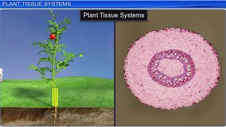
A flowering plant body is made of tissues grouped into tissue systems. Meristematic tissues are actively dividing and are found at specific regions such as root tips, shoot tips, internodes and lateral sides. They give rise to permanent tissues, whose cells lose the ability to divide and become specialised. Permanent tissues are simple when made of one type of cell, such as parenchyma, collenchyma and sclerenchyma, and complex when made of multiple cell types, such as xylem and phloem. The epidermal tissue system forms the protective outer covering with epidermis, stomata, root hairs and trichomes. The ground tissue system includes cortex, endodermis, pericycle, pith and mesophyll. The vascular tissue system consists of xylem and phloem, arranged differently in roots, stems and leaves.
- 1Apical meristem occurs at root and shoot tips and contributes to primary growth.
- 2Intercalary meristem occurs between mature tissues, especially at bases of leaves or internodes in grasses.
- 3Lateral meristem includes vascular cambium and cork cambium.
- 4Epidermis usually has a cuticle in aerial parts, but root epidermis lacks cuticle to absorb water.
- 5Stomata are made of guard cells and regulate transpiration and gaseous exchange.
- 6Ground tissue is all tissue except epidermis and vascular bundles.
- 7Vascular bundles may be radial, conjoint, open or closed depending on organ and plant group.
Meristem Functions
A-I-L: Apical increases length, Intercalary increases internode, Lateral increases largeness or girth.
Simple Tissue Order
PCS: Parenchyma stores, Collenchyma supports flexibly, Sclerenchyma strengthens rigidly.
Grass Regrowth
Grass can regrow after cutting because intercalary meristems remain active near nodes or leaf bases.
Cactus Epidermis
A thick cuticle on aerial epidermis reduces water loss in xerophytic plants.
Calling All Permanent Cells Dead
Parenchyma and collenchyma are living; sclerenchyma is dead at maturity.
Confusing Tissue System with Tissue Type
Xylem is a tissue, while vascular tissue system is the full conducting system containing xylem and phloem.
Forgetting Intercalary Meristem
Intercalary meristem is important in grasses and helps rapid regrowth after grazing or cutting.
A quick functional rule for different meristems.
Variables
apical=Meristem at tips of root and shoot
intercalary=Meristem between mature tissues, common in grasses
lateral=Meristem along sides, responsible for secondary growth
Xylem, Phloem and Vascular Bundles
Overview
Xylem and phloem are complex permanent tissues because each contains more than one cell type. Xylem conducts water and minerals from roots to aerial parts and provides mechanical support. It consists of tracheids, vessels, xylem fibres and xylem parenchyma; except xylem parenchyma, most xylem elements are dead at maturity. Phloem transports organic food, mainly from leaves to other plant parts. It consists of sieve tube elements, companion cells, phloem parenchyma and phloem fibres. Gymnosperms and pteridophytes lack sieve tubes and companion cells but have sieve cells. Vascular bundles may be radial in roots or conjoint in stems and leaves. They may be open if cambium is present or closed if cambium is absent. Exarch xylem is typical of roots, while endarch xylem is typical of stems.
- 1Tracheids and vessels are water-conducting elements of xylem.
- 2Sieve tube elements and companion cells work together in angiosperm phloem.
- 3Phloem parenchyma is absent in most monocots.
- 4Cambium between xylem and phloem makes a vascular bundle open.
- 5Collateral bundle means xylem and phloem are on the same radius, with phloem outside and xylem inside.
- 6Radial bundle means xylem and phloem occur on different radii, alternating with each other.
- 7Protoxylem position is a major clue: exarch in root, endarch in stem.
Xylem vs Phloem
Xylem pulls water from roots upward; Phloem feeds plant parts with food.
Open vs Closed
Open bundle has an open growth option because cambium is present; closed bundle has no cambium.
Exarch-Endarch
Root exits outward: exarch. Stem stores inside: endarch.
Dicot Stem Bundle
A sunflower stem shows open collateral vascular bundles arranged in a ring.
Monocot Stem Bundle
A maize stem has scattered closed collateral vascular bundles without cambium.
Saying All Xylem Cells Are Dead
Xylem parenchyma is living; tracheids, vessels and fibres are dead at maturity.
Forgetting Phloem Parenchyma in Monocots
Phloem parenchyma is generally absent in most monocot stems.
Confusing Radial and Collateral Bundles
Roots have radial bundles with alternating xylem and phloem; stems have conjoint bundles.
The four components of angiosperm xylem.
Variables
tracheids=Elongated water-conducting dead cells
vessels=Tube-like water-conducting elements
xylem fibres=Mechanical support elements
xylem parenchyma=Living storage and lateral conduction cells
The four components of angiosperm phloem.
Variables
sieve tubes=Food-conducting living cells without nucleus at maturity
companion cells=Cells controlling sieve tube activity
phloem parenchyma=Storage and lateral conduction cells
phloem fibres=Dead sclerenchymatous support fibres
Root Anatomy: Dicot Root and Monocot Root
Overview
Root anatomy is identified by epiblema, cortex, endodermis, pericycle and radial vascular bundles. The outermost layer is epiblema or piliferous layer, often bearing root hairs for absorption. Cortex is made of thin-walled parenchyma with intercellular spaces and stores food. The innermost layer of cortex is endodermis, which has Casparian strips made of suberin and regulates water entry into the stele. Pericycle lies just inside endodermis and gives rise to lateral roots and, in dicots, part of vascular cambium and cork cambium during secondary growth. Xylem and phloem are arranged radially and alternately. Xylem is exarch. Dicot roots usually have fewer xylem bundles and small or absent pith, while monocot roots have many xylem bundles and large pith.
- 1Casparian strips block uncontrolled apoplastic movement of water.
- 2Passage cells in endodermis allow water movement near protoxylem points.
- 3Pericycle is important for lateral root origin.
- 4Dicot root commonly has 2 to 6 xylem bundles.
- 5Monocot root is polyarch, with many xylem bundles.
- 6Cambium is absent initially in roots but develops later in dicot root for secondary growth.
- 7Conjunctive tissue lies between xylem and phloem in roots.
Root Layer Order
Every Cool Enduring Plant Visits Pith: Epiblema, Cortex, Endodermis, Pericycle, Vascular tissue, Pith.
Dicot vs Monocot Root
Dicot root has definite few xylem arms; monocot root has many xylem arms and massive pith.
Sunflower Root
Sunflower root is a typical dicot root with fewer xylem bundles and small or absent pith.
Maize Root
Maize root is a monocot root with polyarch xylem and a large central pith.
Calling Root Bundles Conjoint
Root vascular bundles are radial, not conjoint collateral.
Forgetting Exarch Xylem
Roots have protoxylem toward the outside and metaxylem toward the centre.
Confusing Pericycle and Endodermis
Endodermis controls water entry; pericycle gives rise to lateral roots.
The standard outside-to-inside arrangement of root tissues.
Variables
epiblema=Outer absorptive layer with root hairs
endodermis=Innermost cortex layer with Casparian strips
pericycle=Layer giving rise to lateral roots
vascular tissue=Radial xylem and phloem
Stem Anatomy: Dicot Stem and Monocot Stem
Overview
Stem anatomy is recognised by epidermis with cuticle, cortex or ground tissue, vascular bundles and pith. In dicot stems, the epidermis is followed by collenchymatous hypodermis, cortical parenchyma, endodermis, pericycle and vascular bundles arranged in a ring. Each vascular bundle is conjoint, collateral and open because cambium is present between phloem and xylem. Xylem is endarch, meaning protoxylem lies toward the centre. Medullary rays occur between vascular bundles, and pith is well developed. In monocot stems, hypodermis is sclerenchymatous, ground tissue is undifferentiated and vascular bundles are numerous, scattered and closed. Each monocot vascular bundle is surrounded by a sclerenchymatous bundle sheath and contains a protoxylem lacuna. NEET frequently tests ring versus scattered arrangement and open versus closed bundles.
- 1Open vascular bundles allow secondary growth in dicot stems.
- 2Cambium is absent in typical monocot stem vascular bundles.
- 3Dicot stem has distinct cortex, endodermis, pericycle and pith.
- 4Monocot stem lacks clear differentiation into cortex, endodermis, pericycle and pith.
- 5Bundle sheath is prominent around monocot stem vascular bundles.
- 6Endarch xylem is a stem feature, whether dicot or monocot.
- 7Monocot vascular bundles are more numerous near the periphery.
Dicot Stem
Dicot stem has bundles in a disciplined ring and cambium for growth.
Monocot Stem
Monocot stem bundles are many and messy: scattered, closed and with bundle sheath.
Stem Xylem
Stem is endarch: protoxylem is toward the inner end.
Sunflower Stem
Sunflower stem is a classic dicot stem with vascular bundles arranged in a ring and cambium present.
Maize Stem
Maize stem is a classic monocot stem with scattered closed vascular bundles and sclerenchymatous hypodermis.
Calling Monocot Bundles Open
Monocot stem vascular bundles are closed because cambium is absent.
Confusing Dicot Root with Dicot Stem
Dicot root has radial bundles, while dicot stem has ring-arranged conjoint bundles.
Ignoring Protoxylem Lacuna
Protoxylem lacuna is a useful monocot stem identification feature.
Outside-to-inside arrangement of typical dicot stem.
Variables
hypodermis=Collenchymatous support layer in dicot stem
vascular bundles=Ring-arranged open collateral bundles
pith=Central parenchymatous region
A compact rule to distinguish stem from root.
Variables
conjoint=Xylem and phloem present in the same vascular bundle
endarch=Protoxylem lies toward centre
Secondary Growth in Dicot Stem and Root
Overview
Secondary growth is the increase in thickness or girth of plant organs due to activity of lateral meristems. It is common in dicot stems and roots and usually absent in monocots. In dicot stem, vascular cambium forms a continuous cambial ring by joining intrafascicular cambium within vascular bundles and interfascicular cambium between bundles. This cambium cuts secondary xylem toward the inside and secondary phloem toward the outside. More secondary xylem is produced than secondary phloem, leading to wood formation. Cork cambium or phellogen develops in the outer cortical region and produces cork or phellem outward and secondary cortex or phelloderm inward. Together phellem, phellogen and phelloderm form periderm. Annual rings, heartwood, sapwood and bark are key NEET terms from secondary growth.
- 1Intrafascicular cambium is present within vascular bundles of dicot stem.
- 2Interfascicular cambium forms from medullary ray cells between bundles.
- 3Spring wood has wider vessels and lighter colour; autumn wood has narrower vessels and darker colour.
- 4Heartwood is older, non-functional, darker and provides mechanical support.
- 5Sapwood is younger, functional and conducts water.
- 6Bark includes all tissues outside vascular cambium.
- 7Lenticels permit gaseous exchange through periderm.
Periderm
Phellem outside, phellogen middle, phelloderm inside: cork cambium is the generator.
Cambium Direction
Xylem goes in, phloem goes out. Wood is inside.
Heartwood vs Sapwood
Heartwood is hard and central; sapwood supplies sap.
Annual Rings
A tree trunk cross-section shows annual rings formed by alternating spring wood and autumn wood.
Lenticels
Small raised pores on woody stems are lenticels, allowing gaseous exchange through periderm.
Calling Bark Only Cork
Bark includes all tissues outside the vascular cambium, not just cork.
Reversing Cambium Products
Vascular cambium produces secondary xylem inward and secondary phloem outward.
Assuming Monocots Commonly Show Secondary Growth
Typical monocot stems lack vascular cambium and generally do not show normal secondary growth.
Continuous vascular cambium ring forms in dicot stem during secondary growth.
Variables
intrafascicular cambium=Cambium present within vascular bundles
interfascicular cambium=Cambium formed between vascular bundles
Explains how wood and secondary phloem are produced.
Variables
secondary xylem=Wood formed toward inner side
secondary phloem=Food-conducting tissue formed toward outer side
Leaf Anatomy: Dicot Leaf and Monocot Isobilateral Leaf
Overview
Leaf anatomy is adapted for photosynthesis, gaseous exchange and transpiration control. A dicot leaf is usually dorsiventral, meaning the upper and lower surfaces are structurally different. It has upper epidermis with cuticle, lower epidermis with more stomata, and mesophyll differentiated into palisade parenchyma and spongy parenchyma. Palisade cells are chloroplast-rich and perform maximum photosynthesis, while spongy parenchyma has intercellular spaces for gas exchange. Vascular bundles occur in veins and are surrounded by bundle sheath cells; xylem is toward the upper epidermis and phloem toward the lower epidermis. A monocot leaf is isobilateral, with similar upper and lower surfaces, stomata on both sides, undifferentiated mesophyll and bulliform cells on the upper epidermis. Bulliform cells help leaf rolling during water stress.
- 1Palisade parenchyma is the main photosynthetic tissue in dicot leaves.
- 2Spongy parenchyma has large air spaces for gaseous exchange.
- 3Epidermis protects the leaf and cuticle reduces water loss.
- 4Guard cells regulate stomatal opening and closing.
- 5Bundle sheath surrounds vascular bundles and provides support and regulation.
- 6Parallel venation in monocots corresponds with similar vascular bundles along the leaf.
- 7Bulliform cells lose water and become flaccid, causing leaf rolling in grasses.
Dicot Leaf
Dicot has different sides: D for Dorsiventral and Different.
Monocot Leaf
Monocot is iso: equal-looking sides, with bulliform cells like balloons.
Leaf Bundle Orientation
Xylem looks upward for water supply; phloem flows downward toward food transport.
Sunflower Leaf
Sunflower leaf is dorsiventral with palisade mesophyll below the upper epidermis.
Maize Leaf
Maize leaf is isobilateral and has bulliform cells on the upper epidermis.
Calling Monocot Leaf Dorsiventral
Typical monocot leaf is isobilateral, not dorsiventral.
Reversing Xylem and Phloem in Leaf Vein
In a leaf vascular bundle, xylem faces upper epidermis and phloem faces lower epidermis.
Ignoring Bulliform Cells
Bulliform cells are a major monocot leaf clue and help in leaf rolling.
A compact identification rule for dicot leaf anatomy.
Variables
dorsiventral=Upper and lower leaf surfaces differ
palisade mesophyll=Columnar chloroplast-rich cells below upper epidermis
spongy mesophyll=Loosely arranged cells with air spaces
A compact identification rule for monocot leaf anatomy.
Variables
isobilateral=Both leaf surfaces are similar
bulliform cells=Large motor cells that help leaf rolling
undifferentiated mesophyll=Mesophyll not clearly divided into palisade and spongy layers
Epidermis, Stomata, Trichomes, Root Hairs and Bulliform Cells
Overview
The epidermal tissue system forms the outermost covering of the plant body and includes epidermal cells, stomata, root hairs, trichomes and specialised cells such as bulliform cells. Epidermal cells are compactly arranged and usually covered by a waxy cuticle in aerial parts to reduce water loss. Root epidermis, called epiblema, generally lacks cuticle and forms root hairs that increase absorptive surface area. Stomata are pores surrounded by guard cells and sometimes subsidiary cells; they regulate gaseous exchange and transpiration. In dicots, guard cells are kidney-shaped, while in grasses they are dumb-bell shaped. Trichomes on stems may reduce transpiration, provide protection or secrete substances. Bulliform cells in monocot leaves are large, thin-walled cells that help leaves roll during water stress.
- 1Epidermal cells are usually compact with few intercellular spaces.
- 2Root hairs are short-lived but greatly increase absorption.
- 3Subsidiary cells are epidermal cells associated with guard cells.
- 4Stomata may be more on lower epidermis in dicot leaves.
- 5Trichomes may be unicellular or multicellular and can be glandular or non-glandular.
- 6Bulliform cells become flaccid during water stress, causing leaf rolling and reducing transpiration.
- 7Cuticle is generally absent in roots because absorption is required.
Bulliform Cells
Bulliform cells behave like balloons: full means flat leaf, empty means rolled leaf.
Root Hair
Root hairs are for reach: they increase surface area to reach water.
Maize Leaf Rolling
During water stress, maize leaf rolls due to loss of turgidity in bulliform cells.
Stem Trichomes
Trichomes on young stems reduce water loss and may protect against herbivores.
Calling Root Hairs Multicellular
Root hairs are unicellular extensions of epiblema cells.
Forgetting Cuticle Absence in Roots
Roots need absorption, so a thick cuticle is generally absent on epiblema.
Mixing Bulliform Cells with Guard Cells
Guard cells control stomata; bulliform cells help leaf rolling in monocots.
The complete functional unit controlling stomatal movement.
Variables
stomatal pore=Opening for gas exchange
guard cells=Cells that open and close the pore
subsidiary cells=Associated epidermal cells supporting guard cells
Master Dicot vs Monocot Anatomy Comparison
Overview
Dicot and monocot anatomy can be mastered by comparing root, stem and leaf together. Dicot roots usually have fewer xylem bundles, small or absent pith and secondary growth, while monocot roots have polyarch xylem, large pith and no secondary growth. Dicot stems have vascular bundles in a ring, open bundles with cambium, collenchymatous hypodermis and distinct pith. Monocot stems have scattered closed bundles, sclerenchymatous hypodermis, undifferentiated ground tissue and protoxylem lacuna. Dicot leaves are dorsiventral with palisade and spongy mesophyll, while monocot leaves are isobilateral with undifferentiated mesophyll and bulliform cells. NEET identification becomes easy when you first decide the organ, then check vascular bundle pattern, cambium, pith and mesophyll.
- 1The first question for any section is: root, stem or leaf?
- 2Radial bundles indicate root; conjoint bundles indicate stem or leaf.
- 3Ring vascular bundles with cambium indicate dicot stem.
- 4Scattered vascular bundles indicate monocot stem.
- 5Palisade mesophyll indicates dicot leaf.
- 6Bulliform cells indicate monocot leaf.
- 7Large pith with polyarch xylem indicates monocot root, not monocot stem.
Stem Comparison
Dicot stem is Ring and Ready to grow; Monocot stem is Scattered and Sealed.
Root Comparison
Dicot root has few xylem arms; monocot root has many xylem arms and a big pith.
Leaf Comparison
Dicot leaf is different-sided; monocot leaf is mirror-sided.
Diagram ID Example
A section with scattered vascular bundles, each surrounded by bundle sheath, is monocot stem.
Leaf ID Example
A section with bulliform cells on upper epidermis and no palisade-spongy differentiation is monocot leaf.
Using Pith Alone for Identification
Pith helps, but always combine it with vascular arrangement and xylem condition.
Confusing Monocot Root and Monocot Stem
Monocot root has radial bundles in a ring around pith, while monocot stem has scattered conjoint bundles.
Assuming All Dicot Organs Have Cambium Initially
Dicot stem has cambium in vascular bundles, while dicot root develops cambium later during secondary growth.
A fast decision rule for root cross-sections.
Variables
radial=Xylem and phloem alternate on different radii
exarch=Protoxylem toward periphery
pith=Central parenchymatous region
A fast decision rule for stem cross-sections.
Variables
conjoint=Xylem and phloem in same bundle
endarch=Protoxylem toward centre
open=Cambium present
closed=Cambium absent
NEET Identification Strategy, PYQ Concepts and One-Page Revision
Overview
NEET questions from Anatomy of Flowering Plants are usually based on recognition, comparison and labelled structure-function relationships. The fastest strategy is to identify the organ first: root, stem or leaf. Roots have radial vascular bundles and exarch xylem. Stems have conjoint bundles and endarch xylem. Leaves have upper and lower epidermis, mesophyll and vascular bundles with xylem facing upward. Next decide dicot or monocot using bundle number, arrangement, cambium, pith, palisade mesophyll or bulliform cells. For tissue questions, remember cell nature and function: parenchyma is living and storage-based, collenchyma is flexible support, sclerenchyma is dead mechanical support, xylem conducts water and phloem conducts food. For secondary growth, track cambium products and cork cambium derivatives.
- 1Always label xylem and phloem orientation before choosing an answer.
- 2Casparian strips, radial bundles and lateral root origin point toward root anatomy.
- 3Bundle sheath and protoxylem lacuna strongly point toward monocot stem.
- 4Palisade mesophyll is a strong dicot leaf clue.
- 5Bulliform cells are a strong monocot leaf clue.
- 6Annual rings form due to seasonal activity of cambium.
- 7NEET often asks direct NCERT terms such as starch sheath, pericycle, endarch, exarch, phellogen and lenticels.
Root-Stem-Leaf Quick ID
Radial root, ring dicot stem, random monocot stem, palisade dicot leaf, bulliform monocot leaf.
Cambium Products
Vascular cambium: X in, P out. Cork cambium: Cork out, cortex in.
High-Yield Words
Exarch root, endarch stem, bulliform monocot leaf, palisade dicot leaf, phellogen cork cambium.
PYQ-Style Example 1
A section has radial vascular bundles, exarch xylem and large pith. It is a monocot root.
PYQ-Style Example 2
A section has sclerenchymatous hypodermis, scattered vascular bundles and protoxylem lacuna. It is a monocot stem.
PYQ-Style Example 3
A leaf section has palisade cells below upper epidermis and more stomata on lower epidermis. It is a dicot leaf.
Answering Before Identifying Organ
First decide root, stem or leaf. Many wrong answers happen when students directly jump to dicot or monocot.
Ignoring Direction Terms
Exarch, endarch, upper xylem and lower phloem are directional clues and must be read carefully.
Overlooking NCERT Labels
Terms like starch sheath, protoxylem lacuna, conjunctive tissue and bundle sheath are direct NCERT-based clues.
A stepwise method for solving diagram-based NEET questions.
Variables
section=Given anatomical cross-section
organ=Root, stem or leaf
vascular pattern=Radial, ring, scattered or leaf vein
xylem position=Exarch or endarch; upper-facing in leaf
Formula Sheet
10A practical NEET rule to identify whether a section is root, stem, dicot or monocot.
Variables
epidermis type=Epiblema with root hairs, epidermis with cuticle, or leaf epidermis with stomata
vascular bundle arrangement=Radial, ring-like, scattered or leaf vein arrangement
xylem condition=Exarch in roots and endarch in stems
cambium presence=Open vascular bundle if cambium present, closed if absent
All organs of flowering plants are built from these three tissue systems.
Variables
epidermal tissue system=Protective outer covering including epidermis, stomata, trichomes and root hairs
ground tissue system=Cortex, endodermis, pericycle, pith and mesophyll
vascular tissue system=Xylem and phloem for conduction
A quick functional rule for different meristems.
Variables
apical=Meristem at tips of root and shoot
intercalary=Meristem between mature tissues, common in grasses
lateral=Meristem along sides, responsible for secondary growth
Every root, stem and leaf section can be analysed using these three systems.
Variables
epidermal=Outer protective and absorptive layer
ground=Bulk tissue for storage, support and photosynthesis
vascular=Conducting tissue made of xylem and phloem
The four components of angiosperm xylem.
Variables
tracheids=Elongated water-conducting dead cells
vessels=Tube-like water-conducting elements
xylem fibres=Mechanical support elements
xylem parenchyma=Living storage and lateral conduction cells
5 more formulas locked
Sign up free to access all formulas with variables and explanations.
Quick Revision
12 Sign up to accessUnlock 12 Quick Revision Points
Sign up free to access all content, practice PYQs, and get AI explanations.
Learning Videos
6 Sign up to access




Unlock 6 Learning Videos
Sign up free to access all content, practice PYQs, and get AI explanations.
NEET PYQs — Anatomy of Flowering Plants
24 Sign up to accessShowing 3 of 24 questions. Sign up to practice all with answers, explanations, and AI help.
Match List-I with List-II: Choose the correct answer from the options given below :
The main function of bulliform cells in grasses is:
Select the correct pair.
Unlock the full Anatomy of Flowering Plants experience
All diagrams, videos, quick revision, PYQ practice with AI explanations — plus mock tests, flashcards, and a personalised study plan.